Oral Vestibule (Lips Cheeks Teeth Gums) - Oral Cavity Anatomy

Content:
Introduction 0:00
External Structures of the mouth: 01:57
Division of the Oral Cavity 02:49
Oral Vestibule 03:02
Anatomy of the Lips 03:24
Anatomy of the Cheeks 04:07
Anatomy of the Teeth 05:28
Tooth Arrangement: 08:24
Anatomy of the Gums 10:15
Channel membership: / @taimtalksmed
Follow my IG: / taimtalksmed
Donation link: https://www.buymeacoffee.com/taimtalk...
General structures of the digestive system:
Oral Cavity
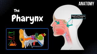
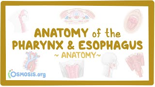
Pharynx
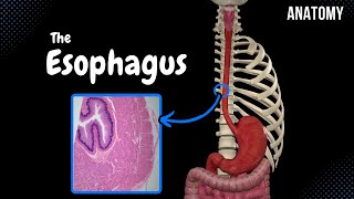
Oesophagus
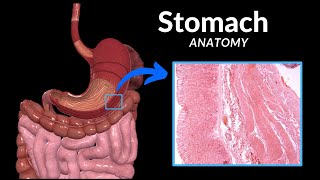
Stomach
Small intestine
Large Intestine
Accessory Organs
Teeth
Tongue
Salivary glands
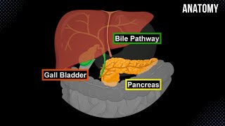
Liver
Pancreas
Gall Bladder
External Structures of the Mouth:
Upper Lip (Labium Superior)
Lower Lip (Labium Inferior)
Oral Angle (Labial Commissure)
Nasolabial Sulcus (Sulcus Nasolabialis)
Philtrum
Mentolabial Sulcus (Sulcus Mentolabialis)
Oral Fissure (Rima Oris)
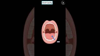
Division of the Oral Cavity:
Oral Vesitibule (Vestibulum Oris)
Oral Cavity Proper (Cavitas Oris Propria)
Oral Vestibule:
External Borders: Lips and Cheeks
Internal Borders: Teeth and Gums
Anatomy of the Lips:
Frenulum of the Upper Lip (Frenulum Labii Superioris)
Frenulum of the Lower Lip (Frenulum Labii Inferioris)
Labial Glands (Glandulae Labialis)
Anatomy of the Cheeks:
Buccinator Muscle (musculus buccinator)
Buccopharyngeal Fascia
Buccal Fat Pad (Bichat's Fat Pad)
Layers of the Skin (cutis)
Tunica Mucosa
Parotid Duct (Ductus Parotideus)
Papilla of the Parotid Duct (Papillae Ductus Parotidei)
Anatomy of the Teeth:
Crown of the tooth (Corona Dentis)
Root of the tooth (Radix Dentis)
Neck of the tooth (Cervix Dentis)
Periodontium
Gomphosis (Dentoalveolar Joint)
Dental Pulp (Pulpa Dentis)
Root Canal (Canalis Radicis Dentis)
Apical Foramen (Foramen Apicis Dentis)
Dentin (Dentinum)
Enamel (Enamelum)
Cement (Cementum)
Milk Teeth (Dentes decidui) 20 teeth, begins at 6th moth of life and ends at 24th month of life
Replacement of milk teeth at age of 612 years
Permanent Teeth (Dentes Permanentes) 32 teeth
Wisdom teeth (dentes serotinus) appears at the age of 1724 years
Tooth Arrangement:
PS! European way of arrangement
First we divide the teeth into 4 equal quadrants
First 2 teeth are called Incisor Teeth (Dentes Invisivi)
Next teeth are Canine Teeth (Dentes Canini)
Premolar Teeth (Dentes Premolares)
Molar Teeth (Dentes Molares)
8*4=32 means we have 32 premanent teeth
Milk Teeth have two incisor teeth, one canine tooth and two molar teeth
Anatomy of the Gums (Gingiva):
Alveolar Mucus Membrane (Covers at the level of root of the tooth. Contains a lot of submucosal Connective tissue)
Gum Proper (attached to the periosteum)
Gingival Papillae (papillae gingivales)
Gingival Margin (margo gingivalis)
Gingival Sulcus (sulcus gingivalis)
Sources used in this video:
Memorix Anatomy 2nd Edition by Hudák Radovan (Author), Kachlík David (Author), Volný Ondřej (Author)
Biorender
University notes and lectures