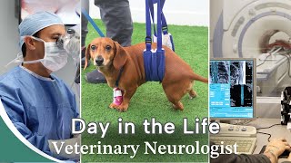
Veterinarian Finds Out Her Dog Has A Brain Tumor

Chesney is an 8yearold, male neutered, Shepherd mix. About 2 weeks prior to us meeting Chesney, his mom noticed that he developed a weak and wobbly gait in his back legs. Over the following days, his front legs also became affected. Chesney’s mom, being a veterinarian, had some testing performed including blood work, xrays, and an ultrasound of his belly. This testing was all normal and did not explain Chesney’s signs. Chesney’s mom began treating him empirically with antibiotics and antinausea medications. Unfortunately, his signs progressed.
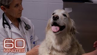
When we met Chesney, he was weak and wobbly in all four limbs, and he had a tendency to lean and fall to his right side. He had a rightsided head tilt and a “drop” in the position of his right eye. Chesney had difficulties knowing where his left front and back legs were in space. Based on his mom’s description of his behavior at home and his examination findings, we were most concerned about a problem affecting Chesney’s balance system.
There are two separate parts of the balance system the middle/inner ear and the back part of the brain (brainstem and cerebellum). In Chesney’s case, we were more concerned about a problem affecting his brain. Chesney’s mom had done a lot of the testing we recommend before doing an MRI. On the day we met Chesney, an MRI of his head was performed. This allowed us to look at both parts of the balance system, as the inner parts of the ears and the brain are both evaluated with this testing.
Chesney’s MRI revealed a mass that appeared to originate from the bones of his skull. This mass was on the left, back part of his skull, and was putting significant pressure on the normal brain inside of the skull. After his MRI, we performed a CT scan of Chesney’s head to get better detail of the mass. Although an MRI is a great diagnostic tool to look at the brain, a CT scan is better for looking at bony structures. Using these two tests together provided us with the ability to better evaluate this mass and determine the best treatment plan. Chesney’s CT confirmed that this mass was originating from the bone.
Based on his clinical signs and the images, we were most concerned about a type of mass called an MLO (multilobular osteochondrosarcoma). These types of tumors originate from the bones of the skull and cause symptoms when they press on the surrounding brain tissue. A sample of the mass would be needed to say that this is the type of tumor Chesney is affected with 100% certainty, but taking a sample of the tissue was considered a risky procedure. Although some MLOs can be treated surgically, we were concerned by the location of Chesney’s tumor posing a higher risk. Radiation therapy is the other treatment option.
While Chesney’s mom considered these options, we started him on a course of antiinflammatory steroids to relieve the swelling that typically surrounds this type of tumor. Chesney is doing better with this medication and, although he is still a bit wobbly, he is learning to navigate these challenges with the help of his loving mom.
Learn more about Vestibular Disease: https://sevneurology.com/petparents/...
#VestibularDisease #dogwithbraintumor #veterinarymedicine