Structure Of Teeth In Humans - Functions Of Teeth In Human Body - Types Of Teeth

In this video we discuss the structure of teeth in humans, the functions of teeth and the different types of teeth. We also cover the tooth numbering system that dentists use, and the 4 quadrants of the oral cavity.
Transcript/notes
The teeth in your mouth are comprised of tiny components that together make up a complex and fascinating structure.
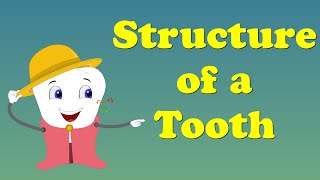
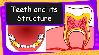
A tooth has 3 main sections, the crown, the neck and the root.
The exposed crown at the top of a tooth is made of enamel, which is comprised mainly of calcium phosphate crystals, and it is the hardest substance in the body. Just below the crown is the neck of the tooth which is surrounded by the gums, and it joins the crown to the root of the tooth.
The root of a tooth fits into what is called an alveolar process of either the maxillae, upper jaw bone or the mandible, lower jaw bone of the skull.
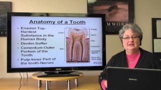
The structure of a tooth consists of the enamel we mentioned earlier, which is a non living tissue, so it can no longer remodel or repair itself. The tooth also has a layer of dentin, which is a harder substance than bone, an inner pulp cavity that is comprised of connective tissue and houses blood vessels and nerves, and a root canal that leads to an opening called apical foramen that opens to surrounding connective tissue.
At the bottom of the tooth, surrounding the root canal and dentin is a hard, mineralized connective tissue called cementum, which I have drawn in on this tooth model, and it helps to connect the tooth to the jawbone. There is a periodontal membrane on the outside of the cementum, which is composed of periodontal ligaments and collagen fibers and houses blood vessels and nerves. This membrane helps to allow the teeth to withstand the stresses of chewing food.
The gums that surround teeth are composed of a top layer of nonkeratinized epithelium and dense irregular connective tissue.
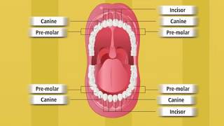
There are 4 types of teeth, incisors, canines, premolars and molars which total 32 permanent teeth in adults 16 maxillary teeth of the upper jaw and 16 mandibular teeth of the lower jaw. This mouth model has only 28 teeth as the back 2 upper and back 2 lower teeth, or wisdom teeth are removed or don’t actually develop in adults many adults. For the purpose of this video and with the help of photoshop, I have added them in.
Dentists use a numbering system for teeth, with 1 thru 16 being teeth of the upper jaw, and 17 thru 32 being of the lower jaw. Teeth 8, 9, 24 and 25 are central incisors, and teeth 7, 10, 23 and 26 are lateral incisors. These teeth have a single root and are designed for cutting into food.
Teeth 6, 11, 22 and 27 are canine teeth, which typically have only one root and they have a pointed tip for puncturing and tearing food.
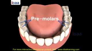
Teeth 5 and 12 are upper 1st premolars and they have 2 roots, and teeth 4 and 13 are upper 2nd premolars which can have 1 or 2 roots. Teeth 21 and 28 are lower 1st premolars, teeth 20 and 29 are lower 2nd premolars and these teeth usually have only one root. Premolars have flat crowns with cusps that allow these teeth to crush and grind food.
Teeth 3, 14, 19, 30 are 1st molars, teeth 2, 15, 18 and 31 are 2nd molars and teeth 1, 16, 17 and 32 are 3rd molars or wisdom teeth. Molars are large teeth that also have flat crowns that are also designed to crush and grind food.
The oral cavity is often divided into 4 quadrants, with each quadrant having 2 incisors, one canine, 2 premolars and 2 or 3 molars.
Timestamps
0:00 The 3 main sections of a tooth (crown, neck, root)
0:37 The structure of a tooth
1:37 The 4 types of teeth
1:51 Numbering system for teeth