Vision: Anatomy and Physiology Animation
Optical and neural components of the eye. Structure of the retina. Fovea and blind spot. Rods and cones, rhodopsin and retinal. Black and white and color vision. Signal transduction in photoreceptor cells, bipolar cells and ganglion cells. Visual projection pathways in the brain. Part of the sensation and perception series sight organ.
Purchase a license to download a nonwatermarked copy of this video here: https://www.alilamedicalmedia.com//g...
Purchase PDF (script of this video + images) here: https://www.alilamedicalmedia.com//g...
©Alila Medical Media. All rights reserved.
Voice by : Marty Henne
Support us on Patreon and get early access to videos and free image downloads: patreon.com/AlilaMedicalMedia
All images/videos by Alila Medical Media are for information purposes ONLY and are NOT intended to replace professional medical advice, diagnosis or treatment.
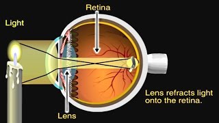
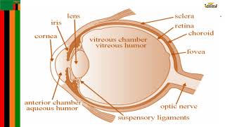
The main optical components are the cornea, the lens, and the iris. The cornea and the lens refract light and focus the image on the retina. The iris acts as an aperture, it controls the amount of light that enters the eye by adjusting the size of the pupil.
The neural components are the retina a lightsensitive tissue lining the inner surface of the eye, and the optic nerve. Light is absorbed by photoreceptor cells in the retina. The optical information is then passed through several cell layers, where it is converted into action potentials and sent, via the optic nerve, to the visual cortex of the brain.
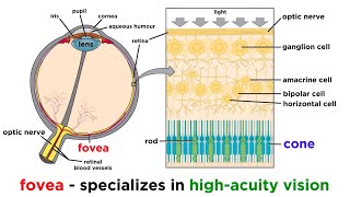
The optic disk, where the optic nerve leaves the eye, has no photoreceptor cells. It corresponds to the blind spot in the visual field.
The major photoreceptor cells of the retina are rods and cones. There are 3 kinds of cones named after the color that they absorb best: red, green and blue. A color is perceived based on proportions of signals coming from these cones. Color blindness occurs when a person lacks a certain kind of cones.
The ability of photoreceptor cells to detect light is due to their lightreceptor molecules, called visual pigments. It’s rhodopsin in rods, and iodopsins in cones. These molecules consist of 2 components: a protein called opsin, and a vitamin Aderivative called retinal.
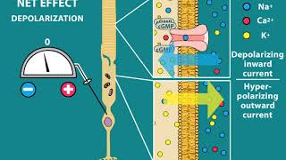
In the dark, there is a socalled dark current in photoreceptor cells. This is due to the presence of cGMP, which permits a constant influx of sodium. The cells are depolarized, they release the neurotransmitter glutamate at the synapse with bipolar cells.
The retinal exists in 2 conformations: cis and trans. In the dark, the cisform is bound to opsin, keeping it inactive. As the retinal absorbs light, it changes to transform and dissociates from the opsin, which now becomes an active enzyme. The enzyme degrades cGMP, sodium channel closes, dark current stops and so does glutamate secretion. The drop in glutamate tells the bipolar cells that light has been absorbed.
On average, each ganglion cell receives signals from over a hundred of rods. This degree of convergence is at the basis of the high sensitivity of rod cells. A dim light produces only a weak signal in a rod, but together, hundreds of these signals converge and become one strong signal acting on a single ganglion cell. However, as the signal comes from a large area of the retina, the image resolution is poor.
The cones have a much lower degree of convergence. The fovea in particular has only cones and no rods, and each cone conveys signal to one ganglion cell. Because one ganglion cell receives input from a very small area of the retina, this setup produces high resolution images. But high resolution comes with low sensitivity, because each cone must be stimulated with a signal strong enough to generate action potentials in the ganglion cell. This also explains why there is no color vision in dim light.
The bipolar cells are firstorder neurons, and ganglion cells are secondorder neurons. The axons of ganglion cells form the optic nerve. The 2 optic nerves from the 2 eyes converge at the optic chiasm. Here, the medial half of nerve fibers from each eye cross to the other side of the brain. Most of the fibers then continue to the thalamus and synapse with thirdorder neurons, whose axons project to the primary visual cortex. Some fibers take a different route: they terminate in the midbrain and are responsible for pupillary light reflex and accommodation reflex, among others.
Note that objects in the left visual field are perceived by the right side of the brain, which also controls motor responses of the body’s left side – the same side as the objects.